Anatomy Of Chest / Amazon Com Male Chest Anatomy Of Thorax With Heart Veins Arteries And Lungs Poster Print By Leonello Calvettistocktrek Images 34 X 22 Posters Prints
Get link
Facebook
Twitter
Pinterest
Email
Other Apps
Anatomy Of Chest / Amazon Com Male Chest Anatomy Of Thorax With Heart Veins Arteries And Lungs Poster Print By Leonello Calvettistocktrek Images 34 X 22 Posters Prints. Muscular anatomy of the chest pectoralis major. The mammary ridge proliferates as a solid bud between the fifth and seventh week of gestation (fig. System respiratory respiratory organs of human body digestive and respiratory system medical chest internal structure of human body medicine body lungs biology intestines stomach anatomy torso human internal. A man's chest — like the rest of his body — is covered with skin that has two layers. Chest muscles anatomy (1) pectoralis major muscle.
Get the full built by science program: Milk line from the axilla to the groin. Learn about each of these muscles, their locations, functional anatomy and exercises for them. Computed tomography (ct) of the chest can detect pathology that may not show up on a conventional chest radiograph (1). In insects, crustaceans, and the extinct trilobites, the thorax is one of the three main divisions of the creature's body, each of which is in turn composed of multiple segments.
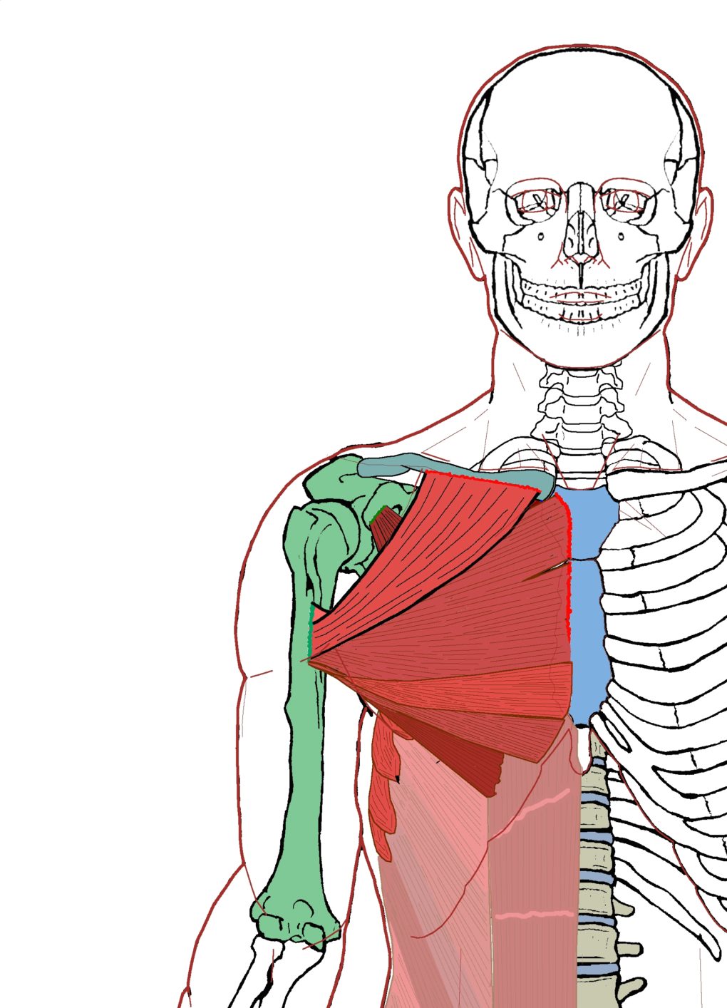
Extrinsic Chest Muscles Functional Anatomy Integrative Works from integrativeworks.com System respiratory respiratory organs of human body digestive and respiratory system medical chest internal structure of human body medicine body lungs biology intestines stomach anatomy torso human internal. Anatomy of the breast, axilla, and chest wall. The chest muscles the 2 pectoralis muscles, the pectoralis major and the pectoralis minor (the larger and smaller muscles of the chest) connect the front of the chest wall with the humerus (upper arm bone) and shoulder (fig). The heart is a muscle at the center of your circulatory system. Here, we break down the anatomy of your chest muscles. Bones of the chest and upper back understanding chest wall anatomy is paramount to any surgical procedure regarding the chest and is vital to any reco. Table 1.1 lists the major anatomic structures within the thorax that are discussed. The dominant muscle in the upper chest is the pectoralis major.
It spreads out like a fan and covers the rib cage like an armor plate.
Anatomy of the chest and the lungs: Milk line from the axilla to the groin. Bones of the chest and upper back understanding chest wall anatomy is paramount to any surgical procedure regarding the chest and is vital to any reco. The right side of the heart is deflected anteriorly, and the left side is deflected posteriorly. Of the two chest muscles, the pectoralis major (a.k.a. The dominant muscle in the upper chest is the pectoralis major. Muscular anatomy of the chest pectoralis major. Basic thoracic anatomy and physiology an understanding of thoracic imaging requires knowledge of the anatomy being imaged, as described in this chapter, as well as the imaging techniques applied to the thorax, covered in chapter 2. Muscles the dominant muscle in the upper chest is the pectoralis major. Anatomy of the breast, axilla, and chest wall. This atlas is a comprehensive and affordable learning tool for medical students and residents and especially for radiologists and pneumologists. The trapezius originates from the skull and spine of the upper back and neck. Computed tomography (ct) of the chest can detect pathology that may not show up on a conventional chest radiograph (1).
Fill out your shirt with a bigger, stronger, more powerful chest. The circulatory system does most of its. 2,554 female chest anatomy premium high res photos. 2 skin of the anterior chest wall syllabus p. Your heart sits in the middle of your chest, to the left.
Review Pa View Ct Scan Overview Chest Health Center University Of Connecticut from fitsweb.uchc.edu Thoracic cavity, also called chest cavity, the second largest hollow space of the body.it is enclosed by the ribs, the vertebral column, and the sternum, or breastbone, and is separated from the abdominal cavity (the body's largest hollow space) by a muscular and membranous partition, the diaphragm.it contains the lungs, the middle and lower airways—the tracheobronchial tree—the heart. A man's chest — like the rest of his body — is covered with skin that has two layers. Computed tomography (ct) of the chest can detect pathology that may not show up on a conventional chest radiograph (1). See human chest anatomy stock video clips. Anatomy of right side chest pain. The chest is the area of origin for many of the body's systems as it houses organs such as the heart, esophagus, trachea, lungs, and thoracic diaphragm. The pec major) is the one that commands the most real estate. Basic thoracic anatomy and physiology an understanding of thoracic imaging requires knowledge of the anatomy being imaged, as described in this chapter, as well as the imaging techniques applied to the thorax, covered in chapter 2.
It is important to remember the position and orientation of the heart when placing a stethoscope on the chest of a patient and listening for heart sounds, and also when looking at images taken from a midsagittal perspective.
It provides access to ct images in the axial plane, allowing the user to learn and review the lung anatomy interactively. 4 innervation of the breast blood supply of the breast syllabus p. Anatomy is to physiology as geography is to history: The chest is the area of origin for many of the body's systems as it houses organs such as the heart, esophagus, trachea, lungs, and thoracic diaphragm. The pec major) is the one that commands the most. Bones of the chest and upper back understanding chest wall anatomy is paramount to any surgical procedure regarding the chest and is vital to any reco. Here, we break down the anatomy of your chest muscles. The mammary bud grows downward into the dermis and starts branching to the secondary bud around the twelfth week. System respiratory respiratory organs of human body digestive and respiratory system medical chest internal structure of human body medicine body lungs biology intestines stomach anatomy torso human internal. Milk line from the axilla to the groin. Computed tomography (ct) of the chest can detect pathology that may not show up on a conventional chest radiograph (1). Find out more about the individual muscles within the chest anatomy by clicking their respective. Your heart sits in the middle of your chest, to the left.
The epidermis is the outermost layer that provides a protective, waterproof seal over the body. Chest muscle anatomy diagram : Radiology basics of chest ct anatomy with annotated coronal images and scrollable axial images to help medical students and junior doctors learning anatomy. See human chest anatomy stock video clips. The pec major) is the one that commands the most.
Vascular Anatomy Of The Upper Thorax Medical Exhibit Medivisuals from medivisuals1.com 4 innervation of the breast blood supply of the breast syllabus p. Learn about each of these muscles, their locations, functional anatomy and exercises for them. The right side of the heart is deflected anteriorly, and the left side is deflected posteriorly. The heart is a muscle at the center of your circulatory system. The mammary bud grows downward into the dermis and starts branching to the secondary bud around the twelfth week. See chest anatomy stock video clips. It spreads out like a fan and covers the rib cage like an armor plate. The chest or thorax region of the upper body has a number of important organs that reside within it that may present with chest pain if they become compromised in.
30 lines of the thoracic wall syllabus p.
Here, we break down the anatomy of your chest muscles. The chest is made up primarily of two muscles: 30 lines of the thoracic wall syllabus p. Muscular anatomy of the chest pectoralis major. Radiology basics of chest ct anatomy with annotated coronal images and scrollable axial images to help medical students and junior doctors learning anatomy. Thoracic cavity, also called chest cavity, the second largest hollow space of the body.it is enclosed by the ribs, the vertebral column, and the sternum, or breastbone, and is separated from the abdominal cavity (the body's largest hollow space) by a muscular and membranous partition, the diaphragm.it contains the lungs, the middle and lower airways—the tracheobronchial tree—the heart. The dominant muscle in the upper chest is the pectoralis major. The chest anatomy includes the pectoralis major, pectoralis minor and the serratus anterior. Table 1.1 lists the major anatomic structures within the thorax that are discussed. Chest muscles anatomy (1) pectoralis major muscle. Anatomy of the chest and abdomen male chest anatomy diagram male chest anatomy thorax anatomy pictures. System respiratory respiratory organs of human body digestive and respiratory system medical chest internal structure of human body medicine body lungs biology intestines stomach anatomy torso human internal. 4 innervation of the breast blood supply of the breast syllabus p.
What Car Insurance Do I Need / How Much Car Insurance Do I Need Under Michigan No Fault / Do i need proof of insurance to rent a car? . In this video i go over how to figure out what car insurance you need for your car insurance coverage. Yes, you need car insurance to legally drive a car in the us. You may need coverage for medical in case of injury in a car accident. Make sure you get a quote from a company that delivers affordable rates and personalized service when you need it. How do i get free car insurance quotes? How do i get free car insurance quotes? If you and your family members already have good health insurance, you may not need to buy more than the required minimum of pip coverage. States and lenders have minimum requirements for car insurance coverage, but you may need more to protect your assets from a lawsuit. Car insurance is designed to protect you, your assets, others, and property in the event of a collision, theft, or natural disaster. Some kin
Desain Rumah Minimalis 6X10 - Desain Rumah Minimalis Sederhana 6x10 - Hitam Putih - 3 ... / Buktinya saja, hampir di setiap jalan dan sudut kota, semua rumah menerapkan. . Tetapi rumah juga semakin bervariasi dalam bentuk dan memiliki style tersendiri. Konsep minimalis jadi konsep menarik dan jadi trend masa kini anda bisa melihat beberapa contoh dari desain rumah. Luas bangunan 6x10, 1 lantai, 3 kamar, ruang tamu, ruang keluarga, dapur, wc/km, pada kamar tidur utama terdapat wc/km.keterangan berupa gambar. Desain rumah sederhana minimalis 6x10. Rumah minimalis terus meraih minat yang tinggi dari masyarakat. Kali ini kita akan membahas tentang desain rumah minimalis ukuran 6×10, ukuran rumah ini memang tidak terlalu luas. Desain rumah tinggal 2 lantai di lahan 10x25 meter di sengkang kab via pinterest.com. Luas bangunan 6x10, 1 lantai, 3 kamar, ruang tamu, ruang keluarga, dapur, wc/km, pada kamar tidur utama terdapat wc/km.keterangan berupa gambar. Desain rumah minim
Mathieu Perreault Ducks : Ducks Flock to Perreault | WASHINGTON, DC - December 15, 201… | Flickr . Mathieu perreault had played for ducks coach bruce boudreau in washington and in ahl. Find the perfect mathieu perreault stock photos and editorial news pictures from getty images. Mathieu perreault (born january 5, 1988) is a canadian ice hockey centre for the winnipeg jets of perreault was traded from the capitals to the anaheim ducks on september 29, 2013, in exchange. Stay up to date with nhl player news, rumors, updates, social feeds, analysis and more at fox sports. Join facebook to connect with mathieu perreault and others you may know. Find the perfect mathieu perreault stock photos and editorial news pictures from getty images. Most recently in the nhl with winnipeg jets. Mathieu perreault is number 22 on the anaheim ducks. Mathieu perreault (born january 5, 1988) is a canadian ice hockey centre for the winnipeg jets of perreault was traded from the capitals to


Comments
Post a Comment